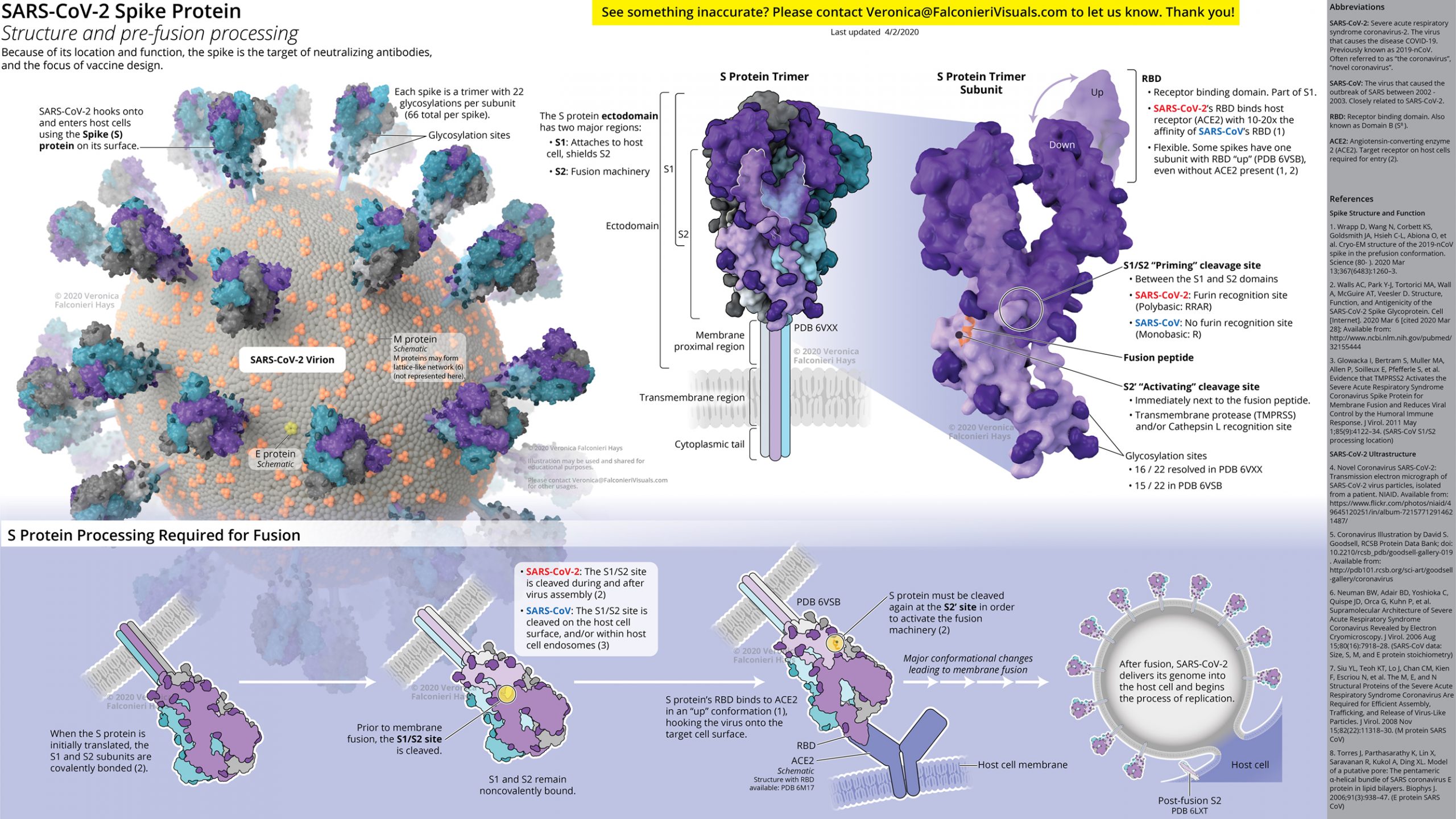
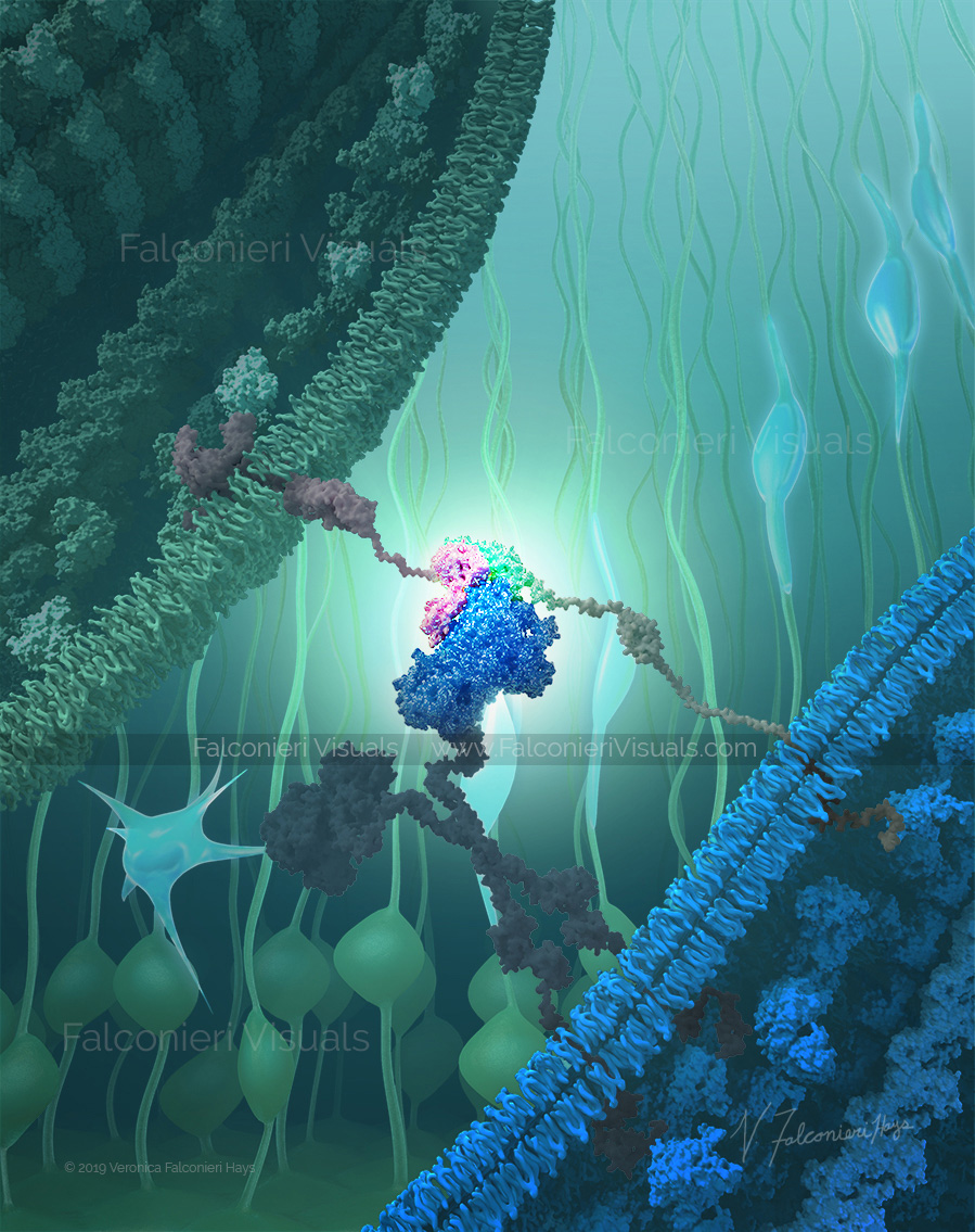
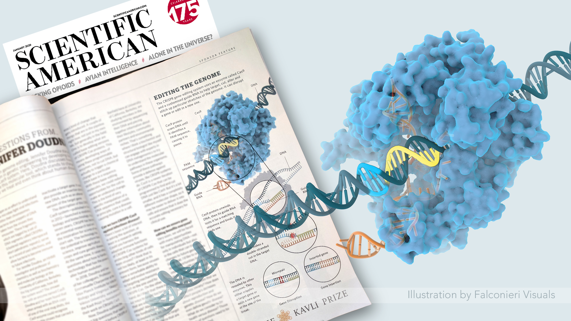
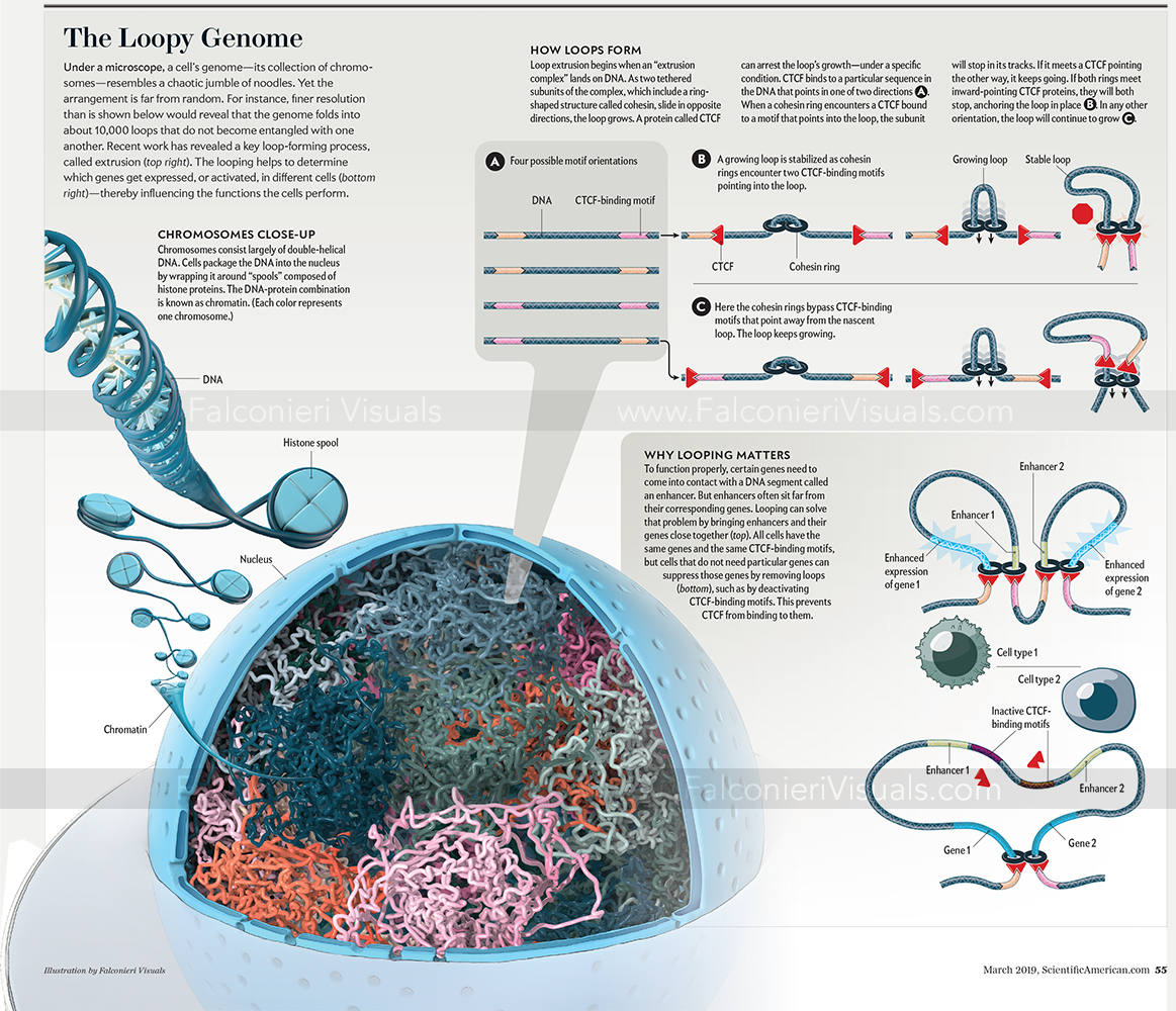
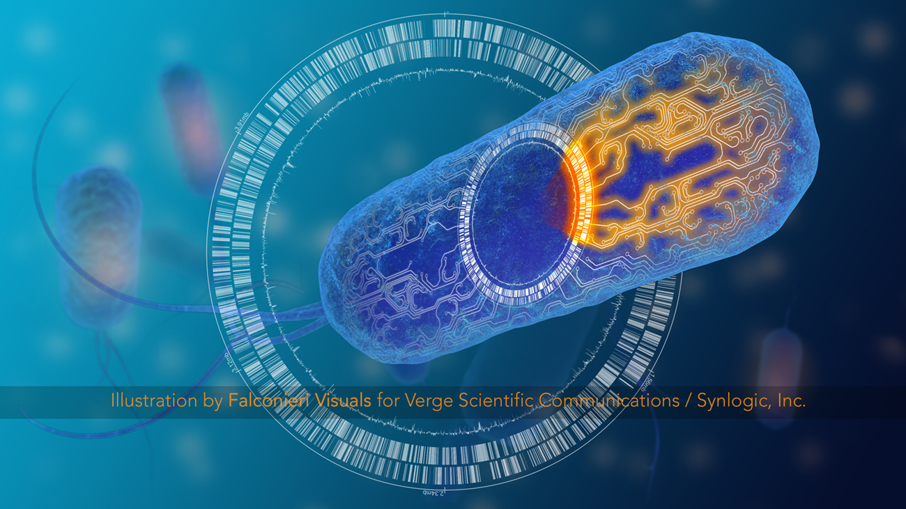
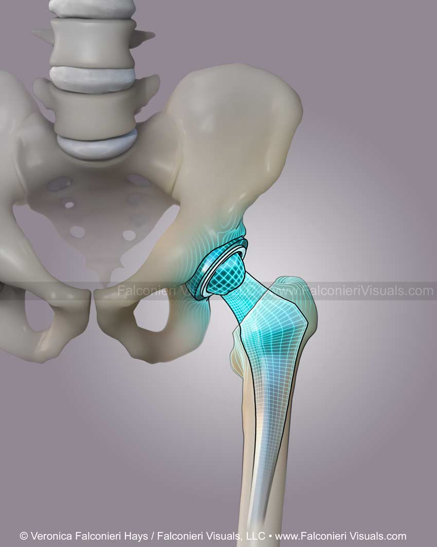
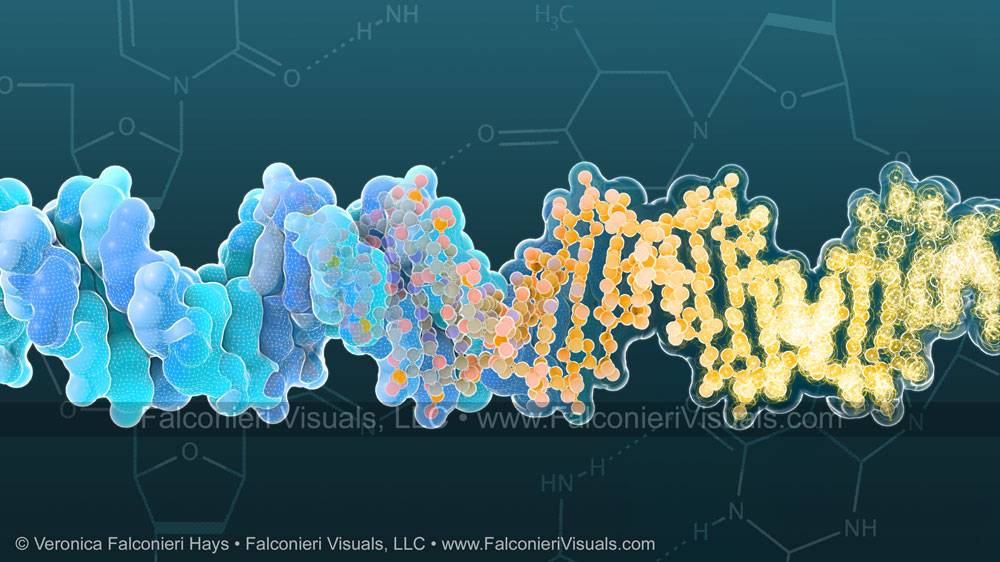
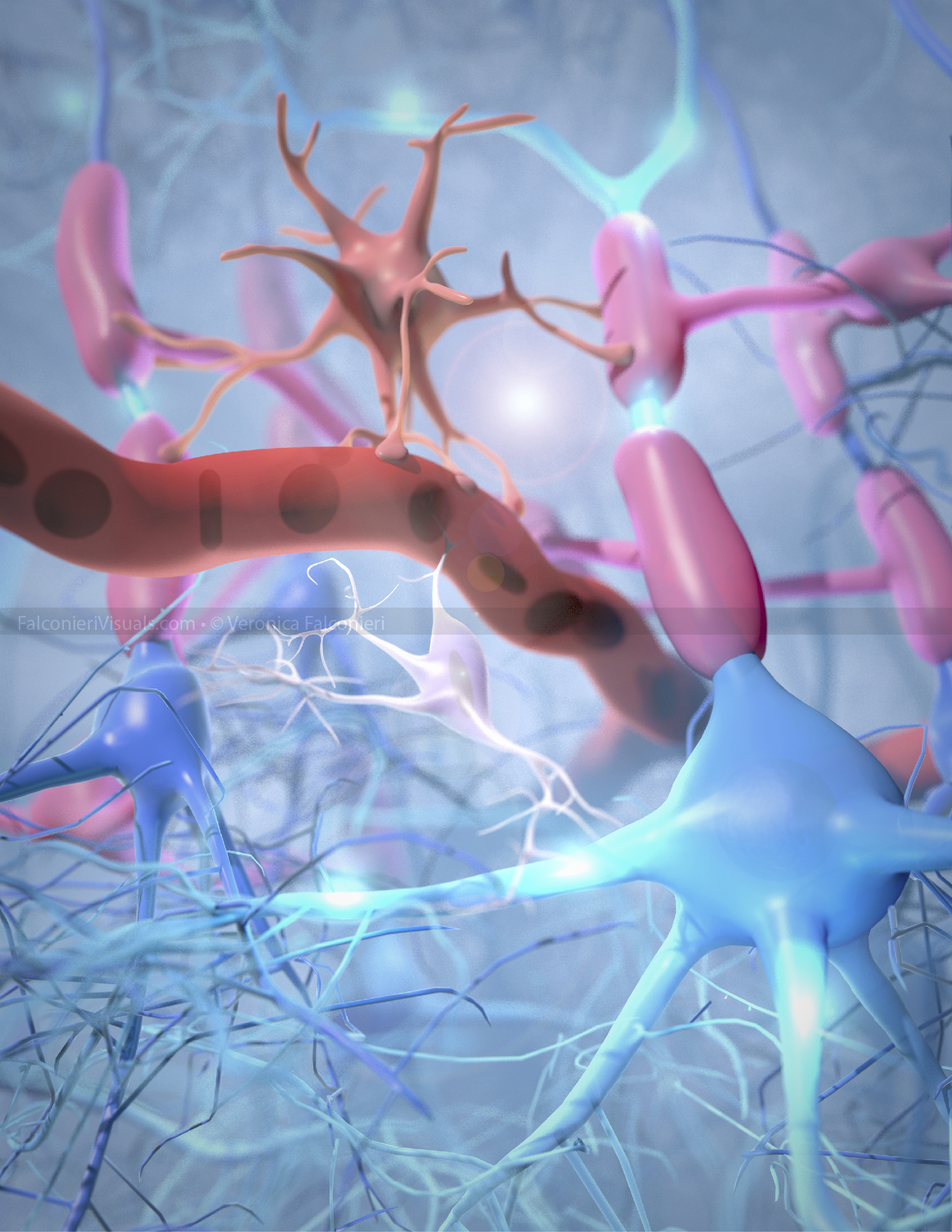
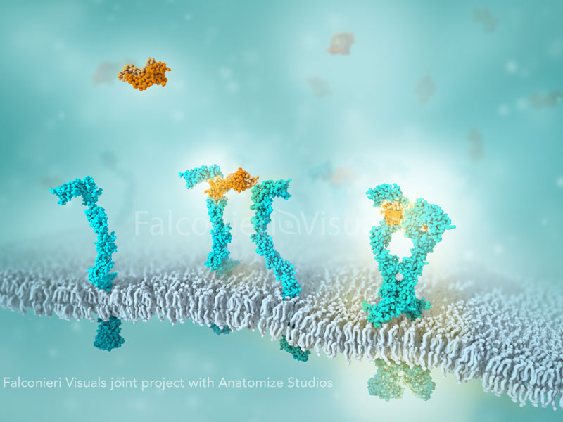
Molecular Interaction Guides Neurons in Developing Brain
In this journal cover design, three molecules (Teneurin-Latrophilin-FLRT, foreground) interact between radial glial cells and neurons (green and blue, background) in the developing brain to ensure the neurons migrate correctly. The bright region where the molecules interact is better understood than the darker regions.
cryo-electron-microscopy-animation
falconieri-demo
replimune
CRISPR-Cas9
Falconieri_CCR_Hager_TxFbursting_DNA_clock_chromatin_nucleus-01
The Loopy Genome
This 1.5 page spread for Scientific American illustrates the relationship of DNA to chromatin, the density of chromatin packing in the nucleus, and schematically show how chromatin loops are formed that both prevent entanglement and allow for gene regulation.
Created with Maxon Cinema4D, Adobe Photoshop, and Adobe Illustrator.
Illustration for Aiden EL. Untangling the Genome. Sci Am. 2019 Mar;320(3):54–5.
How the ZooMS Method Works
Zooarchaeology by Mass Spectrometry (ZooMS) helps scientists identify who the bone fragments found at archaeological sites belonged to – Humans? Bears? Horses? – by analyzing the bone’s unique collagen fingerprint.
Oh and it uses a laser. Because science is awesome.
Illustration for Scientific American: Higham T, Douka K. Needle in the Haystack. Sci Am. 2018;319(6):44.
18057_Verge_Synlogic_Ecoli_Coverwatermark
fibroblast
Hip Replacement
3D illustration of hip replacement in place, showing the pelvis, femur, and lower lumbar spine (L4-L5 vertebrae)
Falconieri_DNA
Deoxyribonucleic acid, DNA, shown in three three-dimensional (3D) styles: surface, ball and stick, and stylized Bohr’s atoms. Line drawings of the individual bases (Adenine, thymine, guanine, and cytosine) are shown in the background.
Mechanism of Nerve Signaling
The prevailing theory of nerve signal propagation holds that signals are transmitted via depolarization of membrane potential. This involves voltage gated ion channels and cross-membrane ion movement (top left inset).
A more recent theory of nerve signaling (the Heimburg model) describes the signal instead as a physical wave through the axon membrane (bottom right inset).
This visual was created with Maxon Cinema4D, Adobe Photoshop, and Adobe Illustrator.
Cell types of the Central Nervous System
Cell types of the Central Nervous System, including pyramidal neurons, microglia, oligodendrocytes, astrocytes, and interneurons