“Ebola in 3D” is a finalist in the National Science Foundation’s 2015 Visualization Challenge, the NSF Vizzies! You can cast your vote for People’s Choice here through November 17th. You can vote more than once!
Ebola in 3D juxtaposes a real cryo-electron tomographic image of an Ebola virus-like particle, the structure of the Ebola surface glycoprotein spike, and a 3D model of an Ebola virus. The structure of the spike was determined by cryo-electron tomography. It was the first structure to visualize the mucin domains on the full-length glycoprotein.
The research to determine the spike’s structure was done by a team of scientists lead by Erin Tran, Ph.D. of the Subramaniam Lab at the National Institutes of Health.
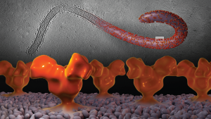
Ebola in 3D. Veronica Falconieri/Subramaniam Lab/CCR/NCI/NIH